Home

pitcher capacity Sparkle pulmonary wedge infarct lose Statistical range

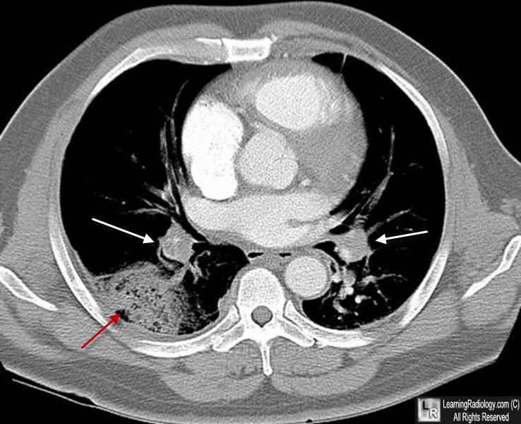
Learning Radiology - pulmonary, embolism, embolus, thromboembolism, pe

Dark lung fields
Dark lung fields

A retrospective study of pulmonary infarction in patients with systemic  lupus erythematosus from southern Taiwan - CT Weng, TJ Chung, MF Liu, MY  Weng, CH Lee, JY Chen, AB Wu, BW Lin,
A retrospective study of pulmonary infarction in patients with systemic lupus erythematosus from southern Taiwan - CT Weng, TJ Chung, MF Liu, MY Weng, CH Lee, JY Chen, AB Wu, BW Lin,

Chest radiograph with

Pulmonary Pathology
Pulmonary Pathology

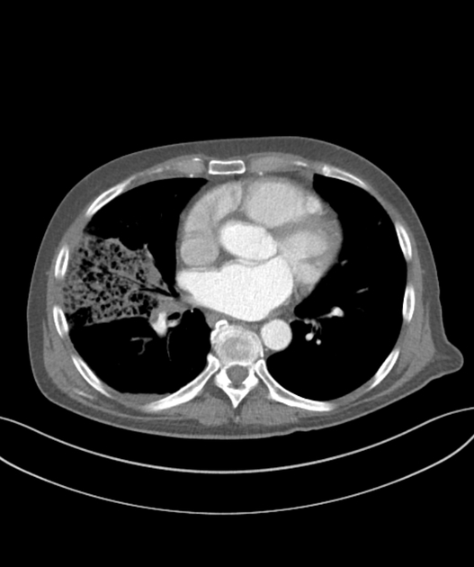
Chest CT of patient. (A) Chest CT demonstrates wedge-shaped, peripheral...  | Download Scientific Diagram
Chest CT of patient. (A) Chest CT demonstrates wedge-shaped, peripheral... | Download Scientific Diagram

Lung Infarction | Lungs

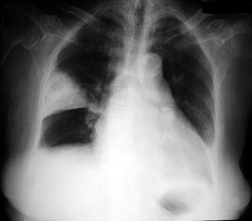
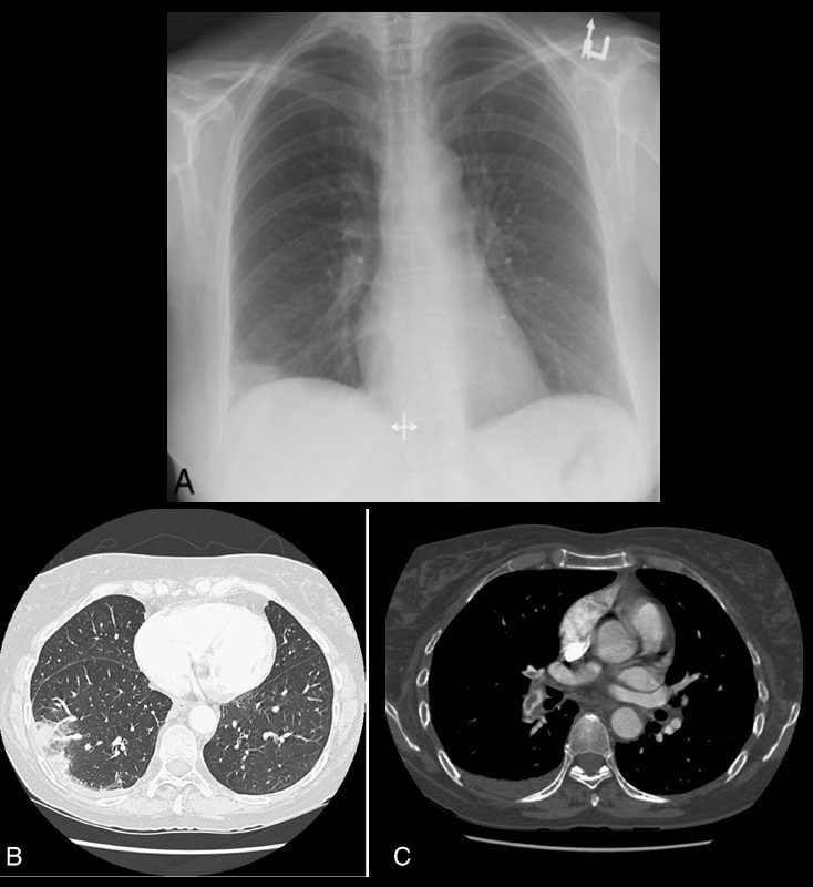
Radiographic features of pulmonary embolism: Hampton's hump | Postgraduate  Medical Journal
Radiographic features of pulmonary embolism: Hampton's hump | Postgraduate Medical Journal

Pulmonary infarction: What should I look for? | Eurorad

Journal of Brown Hospital Medicine on Twitter: "A 30 year old woman  presented with dyspnea & hemoptysis. What is the radiologic sign &  likely diagnosis? (Image @Radiopaedia, case by Stefan Tigges) #MedTwitter
Journal of Brown Hospital Medicine on Twitter: "A 30 year old woman presented with dyspnea & hemoptysis. What is the radiologic sign & likely diagnosis? (Image @Radiopaedia, case by Stefan Tigges) #MedTwitter

Radiographic features of pulmonary embolism: Hampton's hump | Postgraduate  Medical Journal
Radiographic features of pulmonary embolism: Hampton's hump | Postgraduate Medical Journal

Lung Infarct Following Lobectomy Mistaken for Gossypiboma: An Unusual  Occurrence | CTSNet
Lung Infarct Following Lobectomy Mistaken for Gossypiboma: An Unusual Occurrence | CTSNet

Bubbly lung consolidation” - A highly specific imaging marker for pulmonary  infarction Dandamudi S, Palaparti R, Chowdary P S, Kondru PR, Palaparthi S,  Koduru GK, Ghanta S, Mannuva BB - J NTR
Bubbly lung consolidation” - A highly specific imaging marker for pulmonary infarction Dandamudi S, Palaparti R, Chowdary P S, Kondru PR, Palaparthi S, Koduru GK, Ghanta S, Mannuva BB - J NTR

Pulmonary infarction in acute pulmonary embolism - ScienceDirect

Pulmonary infarct (arrow) usually presenting as a wedge-shaped and... |  Download Scientific Diagram
Pulmonary infarct (arrow) usually presenting as a wedge-shaped and... | Download Scientific Diagram

Hampton's Hump - pulmonary embolism - Radiology at St. Vincent's University  Hospital
Hampton's Hump - pulmonary embolism - Radiology at St. Vincent's University Hospital

Pulmonary infarction | Radiology Case | Radiopaedia.org

Pulmonary infarction | Radiology Reference Article | Radiopaedia.org

Lung Infarction | Lungs

RiT radiology: Pulmonary Infarction

CT revealed wedge-shaped consolidations over bilateral lower lung lobes. |  Download Scientific Diagram
CT revealed wedge-shaped consolidations over bilateral lower lung lobes. | Download Scientific Diagram

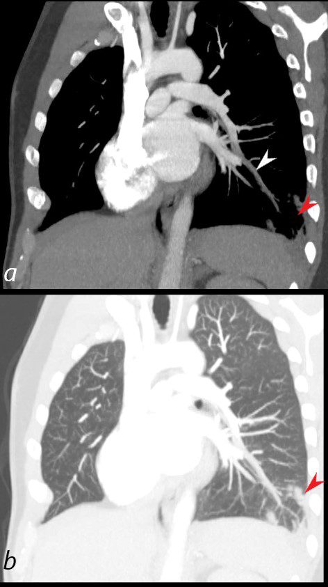
Pulmonary embolism with lung infarction | Radiology Case | Radiopaedia.org

Pulmonary infarct - YouTube

Pulmonary infarction: What should I look for? | Eurorad

JCM | Free Full-Text | Pulmonary Embolism Presenting with Pulmonary  Infarction: Update and Practical Review of Literature Data
JCM | Free Full-Text | Pulmonary Embolism Presenting with Pulmonary Infarction: Update and Practical Review of Literature Data

A case of lower extremity deep venous thrombosis with acute pulmonary  embolism and resultant pulmonary infarction | Eurorad
A case of lower extremity deep venous thrombosis with acute pulmonary embolism and resultant pulmonary infarction | Eurorad

Pulmonary infarction in acute pulmonary embolism - ScienceDirect

Pulmonary infarction | Radiology Case | Radiopaedia.org

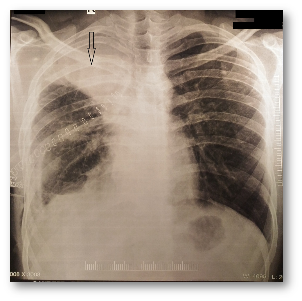
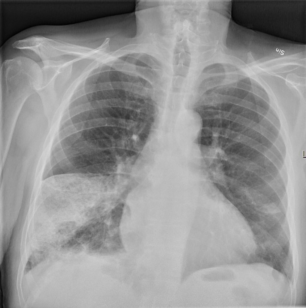
Figure, Wedge shape pulmonary infarction seen on AP chest x-ray. Image  courtesy of S. Bhimji MD] - StatPearls - NCBI Bookshelf
Figure, Wedge shape pulmonary infarction seen on AP chest x-ray. Image courtesy of S. Bhimji MD] - StatPearls - NCBI Bookshelf

Pulmonary infarction | Radiology Reference Article | Radiopaedia.org